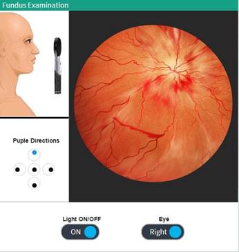
Fundus examination is done as part of a routine physical examination. It is crucial in determining the health of the retina and the vitreous humor. The three basic steps for conducting the fundus examination are Pupil dilation, the wide angle examination of the fundus and zoomed view of fundic lesions/abnormalities.
Ophthalmoscope is simulated in this activity to do fundus examination and to rule out diabetic/hypertensive retinopathy.
|
Simulation Type |
Condition, Using photorealistic graphics |
|
Gender Specific |
No, Common for both gender |
|
Conditions Simulated |
● Fundus exam is Normal ● Hypertensive retinopathy ● Papilledema ● Retinal vein occlusion |
|
Procedure |
● To switch on/off light, click on the 'Light ON/OFF' toggle button. ● A zoomed view of the eye will be shown on the right side. ● To change the pupil directions, click on the respective directions buttons provided under ‘Pupil Directions’. ● Click on the left/right eye toggle button, to examine the desired eye. |