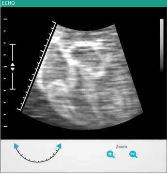
An echocardiogram (also called an ECHO) is a type of ultrasound test that uses high-pitched sound waves that are sent through a device called a transducer. The device picks up echoes of the sound waves as they bounce off the different parts of your heart. The doctor could see how your heart is beating and pumping blood. The doctor can use the images from an echocardiogram to identify various abnormalities in the heart muscle and valves.
|
Simulation Type |
Condition, Using graphics |
|
Gender Specific |
No, Common for both genders |
|
Condition Simulated |
● Normal ● Heart block ● Bradycardia ● Atrial tachycardia ● Ventricular tachycardia ● Ventricular fibrillation ● Atrial fibrillation |
|
Procedure |
● The system will display the patient’s echo on a particular view (like subcostal view) shown in the triangular section. ● The user can pan, tilt & zoom on the image to see other section views. ● Pulse rate, graph, frequency and other values will be displayed along with the echo image. ● The user can note down his observations.
|